
explainer
How to Predict Restriction Digest Fragment Sizes
By Uttam Regmi · Published 2026-07-12 · Updated 2026-08-23 · 6 min read · Fact-checked, sources cited
You’ve cloned something, and before you sequence it you run a quick restriction digest to check it’s right. The gel shows a ladder of bands, but are they the right bands? You don’t have to guess: if you know the sequence, you can predict the exact fragment sizes ahead of time and just confirm the pattern. Here’s how the prediction works.
Step 1: find where the enzyme cuts
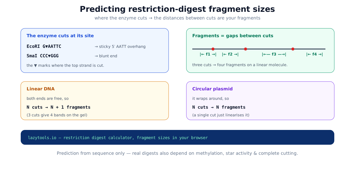
A restriction enzyme binds a specific short recognition sequence and cuts there. EcoRI recognises GAATTC and cuts between the G and the first A (written G^AATTC); SmaI recognises CCCGGG and cuts dead centre (CCC^GGG). To predict a digest you scan the sequence for every occurrence of the site and record the cut position, the base after which the top strand is broken.
Some enzymes have recognition sites with ambiguity: HinfI cuts GANTC, where N is any base. A good calculator expands those IUPAC codes so GAATC, GACTC, GAGTC and GATTC all count as sites.
How often does an enzyme cut?
Before you even scan the sequence, the length of the recognition site tells you roughly how many cuts to expect. If the four bases occurred at random and in equal proportions, a specific restriction site of length n would appear on average once every 4ⁿ bases. That gives a useful rule of thumb:
| Recognition length | Example enzyme | Average spacing | Cuts in a ~5 kb plasmid |
|---|---|---|---|
| 4 bp | HinfI (GANTC) | ~256 bp | ~20 |
| 6 bp | EcoRI (GAATTC) | ~4,096 bp | ~1 |
| 8 bp | NotI (GCGGCCGC) | ~65,536 bp | usually 0 |
That is why 6-bp cutters are the workhorses of routine cloning. They cut a typical plasmid a small, manageable number of times, while 8-bp “rare cutters” like NotI are prized for slicing large genomes into a few big pieces. Real sequences are never perfectly random (GC-content and codon bias skew the odds), so treat these as expectations, not promises: always scan the actual sequence.
Step 2: the gaps between cuts are the fragments
Once you have the cut positions sorted along the sequence, the fragments are simply the distances between consecutive cuts. Take a 54 bp fragment cut at positions 1, 7, 13 and 43:
- start → 1 = 1 bp
- 1 → 7 = 6 bp
- 7 → 13 = 6 bp
- 13 → 43 = 30 bp
- 43 → end (54) = 11 bp
That’s five fragments, 30, 11, 6, 6 and 1 bp, and they add back up to 54. The restriction digest calculator does exactly this for the enzymes you select, and shows each enzyme’s site count so you can spot single-cutters at a glance.
Step 3: linear or circular? It changes the count
This is the part people trip over. The same cut positions give a different number of fragments depending on the DNA’s shape:
- Linear DNA (a PCR product, a gel-purified insert) has two free ends, so N cuts give N+1 fragments. Three cuts → four bands.
- Circular plasmid wraps around with no free ends, so N cuts give exactly N fragments. A single cut doesn’t fragment it at all. It just linearises it into one band at the full plasmid length.
So a plasmid that shows one band after a digest has a single site for that enzyme, a handy way to linearise a vector. Toggle “circular” in the tool when your DNA is a plasmid.
A worked circular example
Say a 5,000 bp plasmid has EcoRI sites at positions 500 and 3,000. Two cuts on a circle give two fragments: from 500 to 3,000 is 2,500 bp, and the piece that wraps around from 3,000 back to 500 (past the origin) is the remaining 2,500 bp. If you had (incorrectly) treated it as linear, you would have predicted three bands, 500, 2,500 and 2,000 bp, and the gel would not match. The topology toggle is the single most common source of a “wrong” prediction.
Sticky ends vs blunt ends
Whether an enzyme leaves a sticky or blunt end depends on where within the site it cuts each strand. EcoRI cuts off-centre and leaves a four-base 5′ AATT overhang; any two EcoRI fragments have complementary overhangs and can be ligated together, which is what makes sticky-end cloning directional and efficient. SmaI cuts straight across the middle and leaves a blunt end, ligatable to any other blunt end, but with no overhang to enforce orientation. Enzymes such as PstI and KpnI cut past the centre on the other strand and leave a 3′ overhang instead.
Here are a few common enzymes and the ends they leave (^ marks the top-strand cut):
| Enzyme | Recognition | Cut | End type |
|---|---|---|---|
| EcoRI | GAATTC | G^AATTC | 5′ overhang (AATT) |
| BamHI | GGATCC | G^GATCC | 5′ overhang (GATC) |
| HindIII | AAGCTT | A^AGCTT | 5′ overhang (AGCT) |
| PstI | CTGCAG | CTGCA^G | 3′ overhang (TGCA) |
| KpnI | GGTACC | GGTAC^C | 3′ overhang (GTAC) |
| SmaI | CCCGGG | CCC^GGG | blunt |
| EcoRV | GATATC | GAT^ATC | blunt |
Overhang compatibility matters for cloning but not for predicting fragment sizes, a 5′ overhang and a blunt cut at the same position produce the same band on a gel. The overhang only becomes relevant when you ligate.
Double digests: pool the cut positions
Cutting with two enzymes at once just means combining their site lists. Suppose a 3,000 bp linear fragment has one EcoRI site at position 1,000 and one BamHI site at position 2,200. Pool the cuts, 1,000 and 2,200, and the linear molecule (two cuts, so three fragments) yields 1,000 bp, 1,200 bp and 800 bp, summing back to 3,000. Double digests are the backbone of restriction mapping because each added enzyme multiplies the information in the banding pattern. The catch is entirely in the wet lab: both enzymes must be active in one shared buffer, or you run them sequentially with a clean-up step in between.
What the prediction can’t tell you
A sequence-only prediction assumes every site is cut, completely and cleanly. Real digests have wrinkles:
- Methylation, Dam (GATC) and Dcm (CCWGG) methylation in most lab E. coli strains blocks some enzymes from cutting overlapping sites.
- Star activity, under high glycerol, wrong buffer or too much enzyme, some enzymes cut relaxed or extra sites.
- Partial digestion, too little enzyme or time leaves some sites uncut, adding larger “partial” bands.
- Double digests, two enzymes need a shared buffer where both are active, or a sequential digest.
Treat the predicted fragments as your expected pattern, then confirm on the gel. For the reverse-complement strand or to check an insert’s orientation, the reverse complement tool pairs well with this one, and everything runs in your browser, so an unpublished construct is never uploaded.
Recognition sites and cut positions here follow standard NEB/REBASE values for 26 common enzymes. The tool predicts a digest from the DNA sequence alone; it does not model methylation, star activity, partial digestion or buffer compatibility. Confirm important results experimentally.
Frequently asked questions
How do I predict restriction digest fragment sizes?
Find every recognition site for your enzyme(s) in the sequence, note where each one cuts, then the fragment sizes are the distances between consecutive cut positions (plus the two ends for linear DNA, or wrapping around for a circular plasmid). A calculator does this automatically from the sequence.
How many fragments will a digest produce?
On a linear molecule, N cuts give N+1 fragments because the two ends are free. On a circular plasmid, N cuts give exactly N fragments because the molecule wraps around, so a single cut just linearises it into one fragment.
What is the difference between a linear and circular digest?
It's the fragment count. Linear DNA (like a PCR product or an isolated fragment) with N cuts gives N+1 pieces; a circular plasmid with N cuts gives N pieces. The same set of cut positions therefore predicts different gels depending on the DNA's topology.
What are sticky ends and blunt ends?
Many enzymes cut the two strands off-centre, leaving a short single-stranded overhang, a 'sticky end' (EcoRI leaves 5' AATT) that base-pairs with any fragment cut by the same enzyme. Others cut straight across, leaving a 'blunt end' (SmaI cuts CCC/GGG). Sticky ends make directional ligation easier.
Why don't my real fragment sizes match the prediction?
A sequence-only prediction assumes every site is cut completely and cleanly. In practice, Dam/Dcm methylation can block some sites, 'star activity' under non-ideal buffer or glycerol conditions can create extra cuts, partial digestion leaves uncut sites, and two enzymes in a double digest need a compatible buffer. Use the prediction as your expected pattern, then confirm on a gel.
How do I do a double digest?
Select both enzymes: the tool finds the sites of each, pools all the cut positions, and computes the fragments from the combined set. Just remember that in the lab a double digest needs a single buffer where both enzymes are active (or sequential digests with a clean-up step).
Can I map an unknown plasmid from its fragment sizes?
Yes, that's restriction mapping. You digest with several enzymes singly and in combination, measure the fragment sizes on a gel, and work backwards to the arrangement of sites. Predicting the fragments from a candidate sequence and matching them to the gel is how you confirm a construct.